")
")
Services
We also offer the development of customer-specific probes.
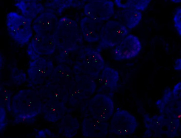
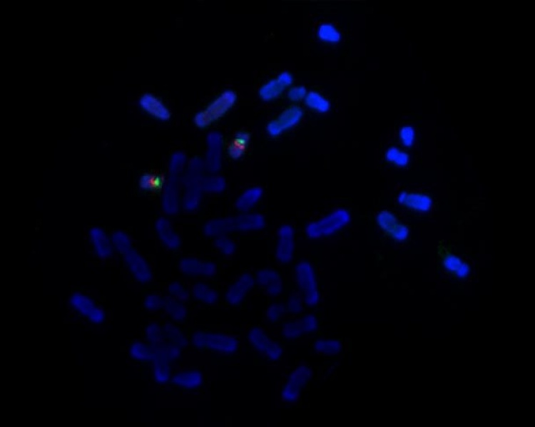
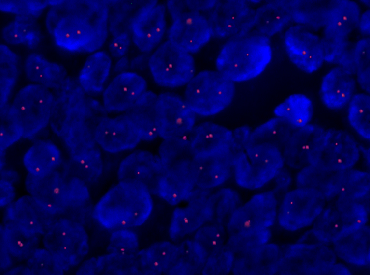
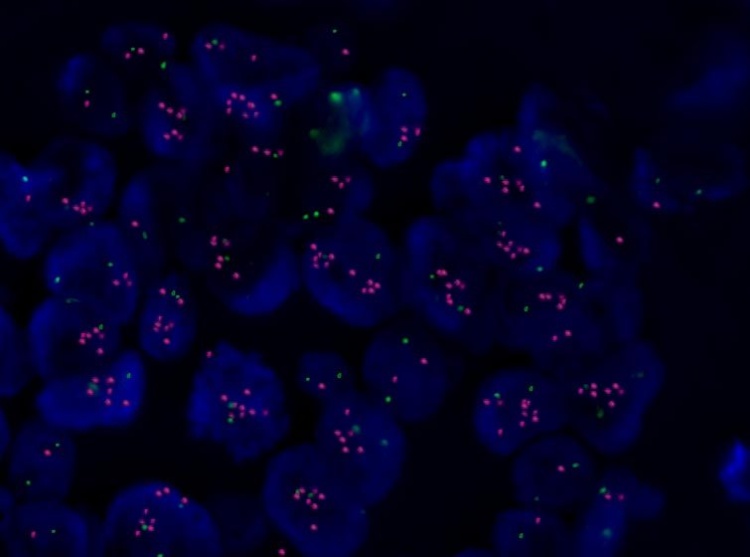
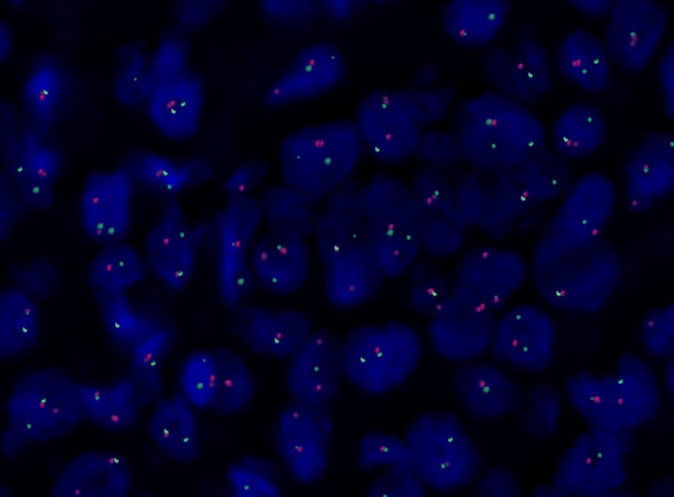
A new fluorescent labeled DNA probe is prepared by purification of specific plasmid DNA of transformed bacterial clone and following direct fluorescent labeling. Bacteria transformed with the appropriate vector are first inoculated into a sterile culture medium and then deposited onto selective agar plates containing the appropriate antibiotic. From each of the agar plates are prepared subclones. After the cultivation, plasmid DNA is isolated. The obtained plasmid DNA is fluorescently labeled using random primers, then precipitated with the addition Cot1 DNA (blocking repetitive sequences) and dissolved in hybridization buffer. The mitotic preparations are used for verifying that the binding region of the probe corresponds to the targeted chromosomal region. Capability (intensity and quality) hybridization probe is screened on various types of specimens (paraffin section, cytospin, mitotic preparation) (see Fig.1).
For a specific request and quotation please contact us.
 / CEP17 (Green) probe with a mitotic preparation") |
 / CEP17 (Green) probe in FFPE tissue.") |
| A | B |
Figure 1: Verification of the binding region of the LSI Her-2 (Orange) / CEP17 (Green) probe with a mitotic preparation (A) and verification of probe ability to hybridizate in FFPE tissue (B).