")
")
LSI Her-2/neu (Orange)/CEP 17 (Green)
« backProbe location

Probe description
The Her-2/neu FISH kit is intended for the determination of Her-2/neu gene amplification in human tissues using fluorescence in situ hybridization (FISH).
The Her-2/neu FISH kit is CE marked and can be used for in vitro diagnostic tests.
The Her-2/neu FISH kit contains two directly labeled fluorescent DNA probes in hybridization buffer. The fluorochrome Orange labeled Her-2/neu probe covers the chromosome 17q11-12 region. The fluorochrome Green labeled chromosome enumeration CEP17 probe covers the chromosome 17p11.1-q11.1 region.
Her-2/neu
The Her-2/neu (Human Epidermal Growth Factor Receptor 2 also known as Neu) gene codes for a 185 kDa transmembrane receptor with tyrosine kinase activity and belongs to the EGF (epidermal growth factor) receptor family. Her-2/neu gene is amplified in 15-30% of breast cancer, and less frequently in other cancers, e. g. lung, pancreatic, ovarian, or stomach cancesr. The Her-2/neu gene amplification is strongly associated with increased disease recurrence and worse prognosis. Breast cancer patients with Her-2/neu gene amplification are treated with monoclonal antibody p185Her-2 trastuzumab (Herceptin) and/or tyrosin kinase inhibitors (lapatinib, Tyverb).
FISH results
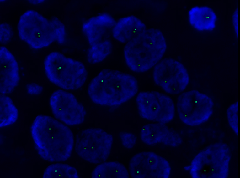
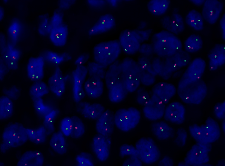
The copy number of Her-2/neu and CEP17 is evaluated in at least 100 cell nuclei in histologically verified section of tissue.
Evaluation of positivity / negativity is carried out according to current recommendations ASCO / CAP.
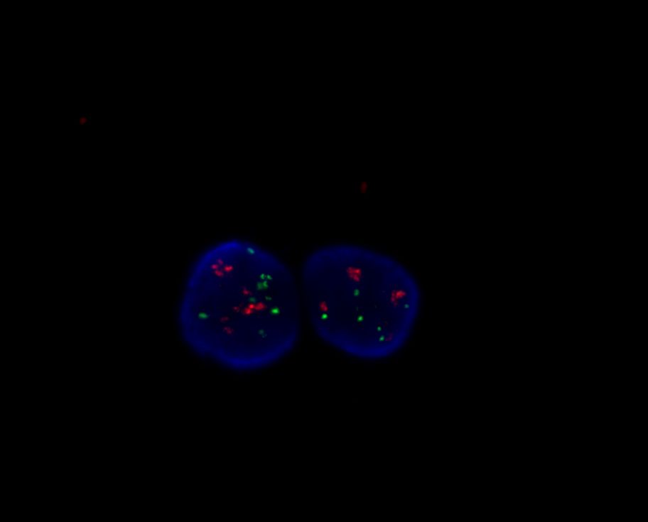
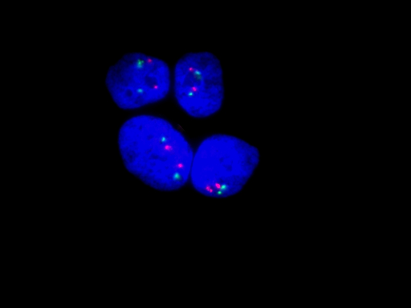
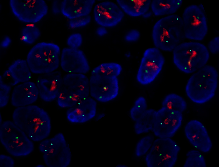
Normally observed in cell two orange signals (Her2/neu) and two green signals (chromosome 17) (Fig. 1a). Figure 1b shows the polyploidy of chromosome 17, accompanied by a higher copy number of Her2/neu gene (pseudo-amplification). True amplification, i.e. a higher number of Her2/neu gene copies with a normal number of chromosome 17, is shown in figure 1c.
/CEP17 (Green) shows 2 orange signals of Her-2/neu gene and 2 green signals of chromosome 17.") |
/CEP17 (Green) shows the polyploidy of chromosome 17 and a higher copy number of Her2/neu gene (pseudo-amplification).") |
/CEP17 (Green) shows 2 green signals of chromosome 17 and higher copy number of Her2/neu gene (amplification).") |
| 1a | 1b | 1c |
Figure 1: Assessment of the copy number of Her-2/neu gene and the copy number of chromosome 17 on FFPE (breast cancer patient tissue). ![]() LSI Her-2/neu
LSI Her-2/neu ![]() CEP17
CEP17
a) Two copies of Her-2/neu genes as well as chromosome 17 in cell (physiological finding).
b) Polyploidia of chromosome 17 with a higher copy number of Her-2/neu gene (pseudo-amplification).
c) Normal copy number of chromosome 17, higher copy number of Her-2/neu gene (true amplification).
Information
|
Probe |
Catalogue number |
Packing size |
Prize |
Instruction for use |
|
LSI Her-2/neu (Orange)/CEP17 (Green) |
IM_001 |
200μl |
upon request |
References
- Wolff, AC, et al.: Recommendations for human epidermal growth factor receptor 2 testing in breast cancer: ASCO / CAP clinical practice guideline update. J Clin Oncol 2013;31(31):3997-40
- Slamon DJ, Clark GM, Wong SG: Human breast cancer: Correrlation of relapse and survival with amplification of the Her-2/neu oncogene. Science 1987; 235: 177-182. [fulltext]
- Perez EA: Her2 as a prognostic, predictive, and therapeutic target in breast cancer, JMCC 1999; 6: 233-240. [fulltext]
- Shak S: Overview of trastuzumab (Herceptin) anti-Her2 monoclonal antibody clinical program in Her2- overexpressing metastatic breast cancer. Semin Oncol 1999; 26: 71-7. [fulltext]
- Ross JS, Fletcher JA: The Her-2/neu oncogene : prognostic factor , predictive factor and target for the therapy. Semin Cancer Biol 1999; 9: 125-138. [fulltext]
- Lebeau A, Deimling D, Kaltz Ch., Sendelhofert, Iff A, Luthardt B, Untch M, Löhrs U: Her-2/neu analysis in archival tissue samples of human breast cancer: comparsion of immunohistochemistry and fluorescence in situ hybridization. J Clin Oncol 2001;19: 354-63. [fulltext]